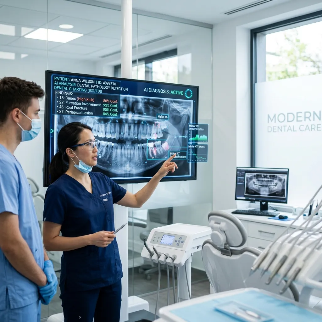
Software that helps read dental images is now part of ordinary practice, and its scope is worth stating plainly. It does three useful things: it flags areas on a radiograph that may be decay, it measures bone levels more consistently than the human eye manages, and it simulates how teeth might move or how an implant might sit in the jaw. That is the honest list. It does not diagnose you and it does not decide your treatment. It is wrong often enough that every flag has to be checked by the dentist sitting with you, and that dentist remains accountable for the decision.
What "AI in dentistry" actually refers to
Nearly all of it is pattern-recognition software. It has been shown large collections of dental images in which experienced dentists already marked what was present — decay, bone loss, a cyst, a fracture line — and learned to mark similar patterns on new images. It is a fast, literal second pair of eyes. It has no understanding of teeth, it has never examined a mouth, and it cannot ask you a question.
Where it genuinely helps
Flagging possible decay on radiographs
Decay between two teeth is easy to miss. It starts where the surfaces touch, out of reach of mirror and light, and on a radiograph may be only a faint shadow a millimetre across. Detection software draws boxes around the regions it considers suspicious, and its value is consistency: it marks the twentieth image of a long day as carefully as the first. It raises questions. The dentist answers them by looking at the tooth, probing the surface, asking about sensitivity and comparing earlier radiographs — the ordinary work of general dentistry.
Measuring bone levels
Gum disease is judged partly by how much bone still supports each tooth, measured on a radiograph from fixed landmarks. By hand that is slow, and two operators rarely produce identical numbers. Software measures the same way every time, which makes comparison across years more meaningful: what matters in gum and periodontal care is not today's figure but whether it has changed. It still says nothing about bleeding, probing depths, mobility or how the gums respond to cleaning — findings that exist only in the mouth.
Treatment simulation and planning
A 3D CBCT scan can be reconstructed so that a planned implant position is checked against the real volume of bone and the position of nerves and the sinus floor before anything is done, which makes planning for dental implants safer. Aligner software stages tooth movement step by step and projects the finished position, a genuinely useful way to discuss braces and aligners before committing.
A simulation is a plan, not a result. Teeth move through living bone at rates that vary, aligners work only while worn, and anatomy sometimes refuses to follow the plan. Treat the image on the screen as the intention.
What it looks like during an appointment
- A radiograph is taken as usual; with a digital RVG sensor it appears on screen in seconds.
- The software marks regions of interest, usually within seconds.
- The dentist reviews the image both with and without the overlay.
- The dentist examines your mouth — looking, probing, tapping, testing response to cold.
- The findings are discussed, including which are certain and which are not.
You will not feel anything different: no extra device in your mouth, no radiation beyond the radiograph being taken anyway, no separate appointment. The step that takes real time is still the conversation.
The limitations, which are the point
False positives are common
Plenty of flagged shadows are not decay. Overlapping tooth surfaces, the margin of an old filling, normal anatomical variation, or a sensor held at a slightly wrong angle all produce marks. If every flag were treated, sound teeth would be drilled. The reasonable response to a borderline flag is often to record it and look again at the next visit.
It also misses things
Early decay beneath an existing crown, a crack running at an unhelpful angle to the beam, the first changes in bone — these can pass unmarked. A clear report is not a clean bill of health.
It cannot do the part that matters most
Software cannot ask what you drink through the day, whether you grind at night, how long pain lingers after cold water, or whether you are pregnant, diabetic or taking blood thinners. It cannot notice that you have avoided one side of your mouth for a month. Diagnosis assembles all of that. An image is one input.
The training images may not resemble you
Much of this software was developed on images from populations whose diet, habits and dental history differ from patients here. Wear from a coarser diet, the tissue changes and staining associated with tobacco and paan, and the patterns left by older restorative work can look unfamiliar to a model and be marked or skipped accordingly. That is a reason for a clinician to read the image themselves.
Accountability does not transfer
No software vendor is answerable for your treatment. If a dentist explains a plan by saying the computer recommended it, ask what they think and why. A good answer refers to the tooth, your symptoms and your history.
Questions worth asking
- Was this flag confirmed by examining the tooth, or only on the image?
- What happens if we record it and review it in six months?
- Can I see the radiograph and have you point it out?
- Is this a measurement, or a projection?
When to see a dentist, whatever the technology
- Pain that has lasted more than a day or two, or that wakes you at night
- Swelling of the gum, cheek or face, or difficulty opening the mouth
- Gums that bleed routinely when you brush
- A tooth that feels loose, or sits high when you bite
- A persistent bad taste or discharge around a tooth
- An ulcer, or a white or red patch, that has not healed in two weeks
None of these should wait on an image being read.
A reasonable expectation
Used sensibly, this software makes records more consistent and surgical planning more precise. Used as a sales argument, it leads to the treatment of findings that were never going to cause trouble. Judge a clinic not on the software it has bought, but on whether the dentist shows you the image, explains the reasoning, and will say when something is uncertain and should be watched.
If you are unsure what a radiograph showed, or you would like a treatment plan looked at again, contact us to arrange an appointment.